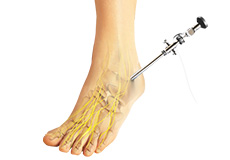
Arthroscopic Surgery
Arthroscopy is a surgical procedure during which the internal structure of a joint is examined for diagnosis and treatment of problems inside the joint. In arthroscopic examination, a small incision is made in the patient’s skin through which pencil-sized instruments that have a small lens and lighting system (arthroscope) are passed. Arthroscope magnifies and illuminates the structures of the joint with the light that is transmitted through fiber optics. It is attached to a television camera and the interior of the joint is seen on the television monitor.
Arthroscopic examination of joints is helpful in diagnosis and treatment of the following conditions:
- Inflammation: Synovitis, the inflammation of the lining of the knee, shoulder, elbow, wrist, or ankle
- Acute or chronic injury: Injuries to the shoulder, knee and wrist joint such as cartilage tears, tendon tears, carpal tunnel syndrome
- Osteoarthritis: A type of arthritis caused by cartilage loss in a joint
- Removal of loose bodies of bone or cartilage that becomes logged within the joint
During arthroscopic surgery, either a general, spinal or local anesthesia will be given depending on the condition. A small incision of the size of a buttonhole is made through which the arthroscope is inserted. Other accessory incisions will be made through which specially designed instruments are inserted. After the procedure is completed arthroscope is removed and incisions are closed. You may be instructed about the incision care, activities to be avoided and exercises to be performed for faster recovery.
Some of the possible complications after arthroscopy include infection, phlebitis (clotting of blood in vein), excessive swelling, bleeding, blood vessel or nerve damage and instrument breakage.
Recovery
It may take several weeks for the puncture wounds to heal and the joint to recover completely. A rehabilitation program may be advised for a speedy recovery of normal joint function. You can resume normal activities within a few days.
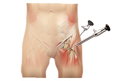
Hip Arthroscopy
Arthroscopy, also referred to as keyhole or minimally invasive surgery, is a procedure in which an arthroscope is inserted into a joint to check for any damage and repair it simultaneously.
An arthroscope is a small, fiber-optic instrument consisting of a lens, light source, and video camera.
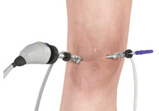
Knee Arthroscopy
The knee joint is one of the most complex joints of the body. The lower end of the thighbone (femur) meets the upper end of the shinbone (tibia) at the knee joint. A small bone called the patella (kneecap) rests on a groove on the front side of the femoral end. A bone of the lower leg (fibula) forms a joint with the shinbone.
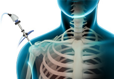
Shoulder Arthroscopy
Arthroscopy is a minimally invasive diagnostic and surgical procedure performed for joint problems. Shoulder arthroscopy is performed using a pencil-sized instrument called an Arthroscope. The arthroscope consists of a light system and camera to project images to a computer screen for your surgeon to view the surgical site.
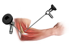
Elbow Arthroscopy
Elbow arthroscopy, also referred to as keyhole or minimally invasive surgery, is performed through tiny incisions to evaluate and treat several elbow conditions.
The Elbow is a complex hinge joint formed by the articulation of three bones - humerus, radius and ulna.
Ankle Arthroscopy
Ankle arthroscopy is a minimally invasive surgical procedure in which an arthroscope, a small, soft, flexible tube with a light and video camera at the end, is inserted into the ankle joint to evaluate and treat a variety of conditions.